 The 7T MRI facility is located in the Life Sciences Centre (see how to find us). It is equipped with a state of the art 7 Tesla Bruker MRI scanner with a 30 cm wide cylindrical bore. We are a user-supported facility which aims to help academic and industrial scientists meet their research goals through the use of MRI technology, particularly for the study of animal models of human disease and disorder.
Our staff can provide support and collaboration with experiment design, MRI technique development, apparatus and sensor development, animal handling, image/data acquisition, and data analysis. Our support infrastructure includes an animal preparation room, in-scan physiological monitoring and maintenance apparatus, computing facilities, access to machining services, and an electronics workshop used primarily for producing custom RF coils (MRI sensors).
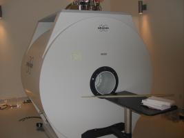
The MRI Scanner
Our primary resource is a Bruker Biospec 7T MRI scanner for NMR imaging and spectroscopy applications. The compatible specimen size is dependent on the size of the gradient coils chosen for the experiment. For typical applications gradients coils of 12 cm or 20 cm inner diameter are used.
- 7.05 Tesla magnetic field at magnet isocentre
- Actively shielded magnet (reduced stray field)
- 4 receiver channels and parallel imaging capabilities
- Scan gating using respiration or ECG signal
- 3 interchangeable gradient sets
- i.d. = 20 cm, max gradient = 200 mT/m (shielded)
- i.d. = 12 cm, max gradient = 400 mT/m (shielded)
- i.d. = 6 cm, max gradient = 1000 mT/m (unshielded)
- Preemphasis unit for eddy current compensation
- Multi-nuclear NMR capability: 1H, 31P, 19F, 13C
- Modular software development: pulse programs, methods, macros
- Commercially available volume RF coils
- i.d. = 15 cm
- i.d. = 6 cm, imaging length = 8 cm
- i.d. = 3.5 cm
Animal care equipment and preparation room
Our facility is equipped with an animal preparation room that is used to prepare an animal for the MRI experiment as well as to allow researchers to perform other procedures and small surgeries. Maintenance of the animal during the MRI experiment is supported by in-scan anesthesia, monitoring and temperature control.
- In-scan anesthesia (primarily isofluorane) and exhaust
- Body temperature control (circulating heated water bath)
- Monitoring and recording of vital signs (respiration, rectal temperature, ECG)
- Tools/equipment for small surgeries (surgical instruments, stereotactic frame, stereomicroscope)
- Remote injection setup and power injector for in-scan i.v. perfusion
- Fume hood (sample preparations and specimen preservation)
- Refrigerator and freezer
- Animal weigh scale and analytical balance
- Grass recorder/stimulator
- Small animal ventilator and intubation kits
RF lab/machining workshop
Our workshop primarily provides infrastructure for the construction of custom RF coils, which are often necessary to optimize the data quality for a given MRI application. We also have some basic machining tools for making experimental apparatus such as scanning platforms. Most of the machining work is either outsourced or performed by Centre staff at other machine shops around the campus.
- Network analyzer (up to 1.5 GHz frequency)
- Power Meter and loads
- Soldering irons and related tools
- Circuit board etching equipment
- Drill press and bandsaw
- Electromagnetic simulation program (Remcom XFDTD)
- Supplies for MRI phantom preparation
Computing facilities
In addition to the console computer controlling the MRI scanner, we have several computers reserved for student use and data analysis.
- Bruker Paravision acquisition and data analysis software
- MATLAB and IGOR Pro programming environments
- Tape drive for backing up console computer
- Mass storage unit for archiving user data
- CD/DVD burners
Capabilities
MRI techniques that are available include the following:
- High-resolution anatomic imaging of small animals or biological specimens
- Diffusion-weighted or diffusion tensor imaging
- Relaxivity (T1, T2, T2*) mapping
- Perfusion MRI
- Functional MRI
- MR angiography
- Cardiac MRI
- NMR spectroscopy (localized and non-localized)
- Chemical shift imaging
- Parallel imaging techniques
Current Applications
Examples of current applications (please see the Approved Projects section for more details):
- High-resolution morphological imaging
- Mouse models of tumour biology and angiogenesis
- perfusion MRI and Vessel Size Index MRI
- diffusion-weighted MRI
- 31P spectroscopy for pH measurement and metabolite concentrations
- 19F spectroscopy for monitoring chemotherapy drug uptake
- Rat models of spinal cord injury
- Morphological imaging of injury pattern
- T2 measurements (myelin water fraction mapping)
- Diffusion Tensor imaging
- Tract-labeling – Manganese-enhanced MRI
- Non-imaging measurement of lean-to-fat mass ratio
- Imaging of Iron-oxide labeled cells in vivo
- Rat brain/heart spectroscopy for measurement of metabolites
- Multimodal MRI-PET imaging
For other possible applications you may want to view the Bruker website.